POCUS Ultrasound: Guide and Information
In clinical practice, POCUS (Point-of-Care Ultrasound) has become an essential assessment tool: fast, portable, and directly integrated into the patient’s physical examination. Performed at the point of care, this non-invasive imaging technique provides immediate, targeted visualization of anatomical structures or potential abnormalities, thereby enhancing diagnostic accuracy and medical responsiveness.
POCUS finds its place in a wide range of settings: emergency departments, general practice offices, primary care facilities, operating rooms, intensive care units, ambulances, and even field medicine.
This guide presents its principles of use, how it works, its clinical applications, and the conditions for implementing this technology, which has become indispensable in modern medical practice.

What is POCUS Ultrasound?
POCUS (Point-of-Care Ultrasound) is a targeted clinical ultrasound performed directly at the patient’s bedside, without going through the imaging department. It is not intended to replace a full ultrasound performed by a radiologist, but rather to quickly answer a specific clinical question as part of the physical examination.
Designed for immediate use at the point of care, this technique relies on portable or ultraportable devices suited to everyday clinical practice. After specific training, the practitioner can obtain in seconds a focused visualization to assess:
The presence or absence of fluid collections (pleural, pericardial, abdominal, etc.)
The function or dynamics of an organ (heart, bladder, gallbladder…)
Direct or indirect signs of acute pathology (pneumothorax, urinary retention, cholecystitis, vascular abnormalities…)
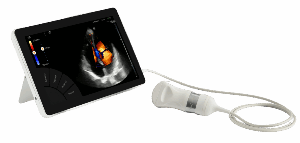
Visualization of a heart on the T-Lite PRO ultraportable Sonoscanner ultrasound device
Today, POCUS ultrasound is used across multiple disciplines: general practice, hospital and prehospital emergency medicine, anesthesia and intensive care, internal medicine, and critical care. It allows clinicians to quickly guide a diagnosis, assist with invasive procedures (such as punctures, drainages, or venous access), or confirm a clinical hypothesis during a focused examination.
How does this technique work?
POCUS ultrasound works by emitting ultrasound waves from a probe applied to the patient’s skin with a conductive gel. The waves are reflected by different tissues, producing a real-time dynamic image that the clinician can interpret immediately or record for later analysis. This technique allows visualization of internal organs such as the heart, liver, or kidneys, detection of pericardial, pleural, or abdominal effusions, assessment of the pleural line and lung ventilation, and identification of abnormalities like pulmonary edema.
The equipment is designed for mobile clinical settings. Using a high-performance portable ultrasound device provides HD image quality while ensuring ergonomics, battery life, and ease of transport.
POCUS is suitable for challenging environments: intensive care units, mobile units, ambulances, hospital departments, or even veterinary medicine. Ultrasound examinations are often guided by standardized protocols, such as FAST, to ensure speed and reliability of the procedure.
How does POCUS change medical practice?
POCUS ultrasound profoundly transforms the way clinicians perform the physical examination. Integrated from the very beginning of a consultation, this rapid and targeted technique allows abnormalities to be identified without moving the patient outside their care setting. It facilitates the assessment of acute conditions, enhances the interpretation of physical signs, and accurately guides invasive procedures or technical interventions.
In emergency medicine, for example, POCUS quickly directs patient management in cases of internal bleeding, pulmonary edema, or abdominal pain.
In general practice, POCUS enables clinicians to go beyond simple palpation, rapidly identify urgent situations, and optimally guide patient care.
Thus, POCUS has become an essential tool for modern, efficient, and structured clinical practice.
In which clinical situations should POCUS ultrasound be used?
This type of POCUS ultrasound is applicable in numerous clinical situations where rapid response and immediate visualization are critical. In emergency medicine, for example, it can be used to detect an abdominal effusion, assess unexplained chest pain, or confirm the presence of pulmonary edema.
In anesthesia, it is used for ultrasound-guided procedures, such as punctures or regional injections.
General practitioners can also use POCUS to check for the absence of deep vein thrombosis, look for fluid collections, or examine a painful gallbladder, thereby improving diagnostic accuracy directly in the office.
In all of these contexts, the quality of professional medical equipment determines the image accuracy, ease of use, and suitability for each specialty: emergency care, primary care, intensive care, or outpatient settings.

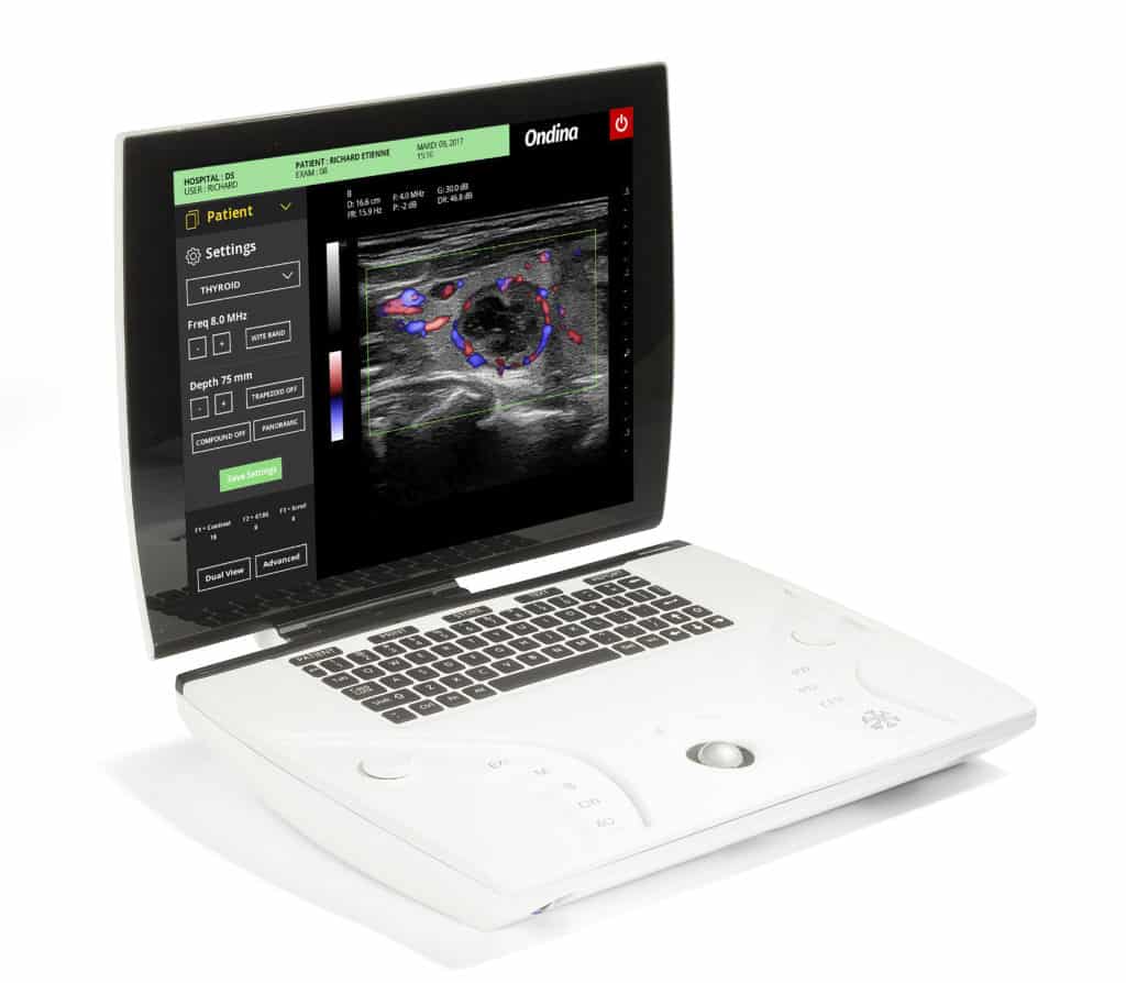
Visualization of a thyroid on the T-Lite PRO portable Sonoscanner ultrasound device
Why the right equipment matters?
The performance of a POCUS ultrasound examination directly depends on the quality of the device used. A portable and reliable system, designed for specific clinical use, ensures quick and precise visualization of internal structures.
High-quality equipment provides a clear image, even under variable lighting conditions or when the patient is moving. Its maneuverability, ergonomics, and lightweight probe significantly impact the clinician’s comfort and the reliability of the results.
In complex environments such as intensive care units, mobile units, or outpatient services, choosing the right equipment enables immediate and optimal patient care, saving valuable time.
Explore further
In clinical practice, POCUS has become a reference ultrasound tool that should be integrated methodically. To go further: