Echogenicity: Definition, Guide, and Best Practices
Echogenicity lies at the heart of ultrasound imaging. This article explores its various aspects and highlights the importance of considering this factor when interpreting medical examinations.
Definition of Echogenicity
Echogenicity refers to a tissue’s ability to reflect a portion of the ultrasound waves emitted by the transducer. These variations in echogenicity depend on how tissues either transmit or reflect sound waves. This property is known as acoustic impedance, which simply describes the tissue’s capacity to resist or allow the passage of ultrasound waves.
As a result, the images produced display varying levels of brightness. Some areas appear whiter or darker on the screen, depending on how the tissues reflect or transmit the ultrasound waves.
The Different Levels of Echogenicity
Professionals identify four main levels of echogenicity, which help differentiate tissues and refine diagnostic analysis.
Anechoic Echogenicity
An anechoic area appears black on the screen because it does not reflect ultrasound waves. It is characteristic of homogeneous fluid-filled structures, such as a cyst or the gallbladder.
Hypoechoic Echogenicity
A hypoechoic structure appears darker than the surrounding tissues. This indicates a low reflection of ultrasound waves, typical of muscles or certain parenchymal organs.
Isoechoic Echogenicity
An isoechoic area reflects ultrasound waves similarly to the surrounding tissues, making it more subtle to identify. Careful attention is required to distinguish isoechoic lesions from healthy tissue.
Hyperechoic Echogenicity
A hyperechoic region appears brighter, sometimes even white, on the image. This results from a strong reflection of ultrasound waves, typically seen in calcifications, fibrotic tissue, or bony structures.
Role of Echogenicity in Ultrasound Diagnosis
Echogenicity helps physicians differentiate tissues and make a diagnosis in medical imaging. It allows them to determine whether an area is normal or if there is an abnormality, such as a nodule or a cyst.
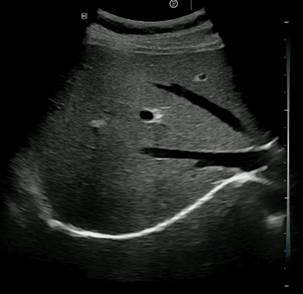
For example, a normal liver has homogeneous echogenicity, neither too bright nor too dark. When a nodule appears hyperechoic (brighter) compared to the rest of the liver, it reflects ultrasound waves more strongly. Some of these nodules are benign (such as hemangiomas), but an accurate diagnosis is necessary.
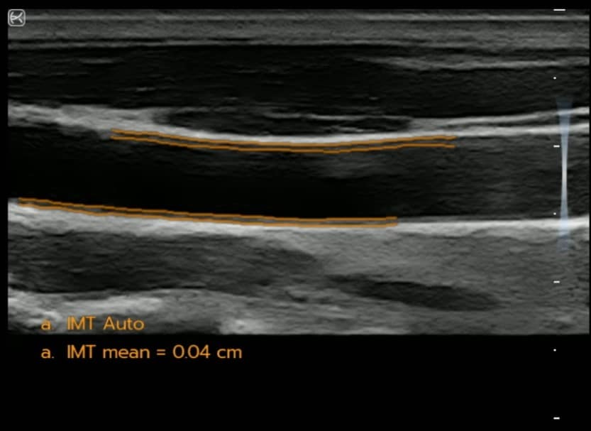
Echogenicity is also used to identify blood vessels and monitor vascular pathologies with Doppler ultrasound. These image contrasts are essential for making a precise diagnosis.

Automatic Measurement of Intima-Media Thickness on the Sonoscanner PRO Range


